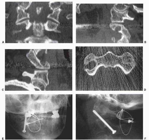

Type III complex odontoid fracture participants were further categorized into two groups, based on treatment received (halo/hard collar) and other inclusion criteria mentioned in the methods section above. Participants were characterized by the type of Type III odontoid fracture (simple/complex), and Chi-square and t-tests/Kruskal–Wallis tests were performed as appropriate to compare the two groups. Univariate descriptive statistics of the study sample were summarized. The maintenance of reduction was defined as <2 mm of change in displacement or 5° of angulation in comparison to initial CT scan. Union was defined as bridging bony consolidation on CT or bony healing of the fracture lines on X-rays in addition to the lack of motion at the fracture site on dynamic radiographs. Stability was defined as the absence of nonphysiologic motion of C1–C2 on dynamic imaging with atlantodens interval >3 mm. Any significant worsening of alignment or displacement prompted recommendations for surgical fusion.įracture stability and union were determined independently by two different authors at the time of discontinuation of immobilization. Displacement was defined as the anterior translation of the odontoid fracture fragment in relation to the C2 or C3 vertebral body.

The mean value of displacement and angulation was utilized for analysis. Fracture deformity was measured off initial CT scans and on final follow-up radiographs with notation of displacement and angulation by two authors. All patients in the study were followed at periodic intervals (1–4 weeks) after discharge from the hospital with upright anteroposterior and lateral radiographs. Patients were treated by both orthopedic and neurological surgeons based on the preferences of the treating surgeon. (a) coronal (b) mid-sagittal computed tomography
ODONTOID FRACTURE COMPLICATIONS TRIAL
Patients that underwent early surgery without a trial of nonoperative management or those lost to follow-up were not included in this study.Įxample of simple Type III odontoid fracture with extension into bilateral superior articular facets. Inclusion criteria for the study were complex Type III fractures that were treated nonoperatively in either a hard collar orthosis (Aspen Collar ®) or halo vest. All other Type III fractures were classified as simple fractures. Fractures with lateral mass comminution >50% or with secondary fractures lines into the pars interarticularis or vertebral body were classified as “complex fractures”. Type III odontoid fractures were divided into two categories. Information regarding patient characteristics including age, mechanism of injury, neurologic status, and other associated injuries (spine, chest, and abdomen) were recorded for all patients with acute Type III odontoid fractures. Fracture morphology was delineated by computed tomography (CT) and classified according to the definition of Type III fracture according to Anderson and D'Alonzo. Institutional review board approval was obtained. Acute C2 (axis) fractures were identified from International Statistical Classification of Diseases-9 coding from 2008 to 2015. The purpose of this study is to separately evaluate treatment outcomes of complex Type III fractures with external immobilization (halo vest and hard collar orthosis).Ī single-institution, retrospective cohort study was conducted to evaluate the outcome of conservative treatment of complex Type III odontoid fractures. High-energy fractures with comminution of the lateral mass and secondary fracture lines extending into the pars interarticularis or vertebral body have historically been studied together with low-energy simple fractures in the elderly. Current studies recommend nonsurgical management of Type III odontoid fractures with union rates of 85%–100% with external immobilization.Ī considerable amount of heterogeneity exists among Type III fractures. In general, the Type III fracture is believed to have high healing potential due to large fracture surface area through cancellous bone. Type III fractures extend into the vertebral body and account of 39% of all odontoid fractures. Type II fractures occur at the junction of the dens and the C2 vertebral body. Type I fractures occur at the proximal tip of the dens from an avulsion injury. The Anderson and D'Alonzo classification is the most commonly utilized classification system. The great majority of odontoid fractures occur in the elderly population from a low-energy mechanism with a smaller contribution from high-energy accidents in the young. The incidence of odontoid fractures varies between age groups and is generally believed to account for approximately 20% of all cervical spine injuries.


 0 kommentar(er)
